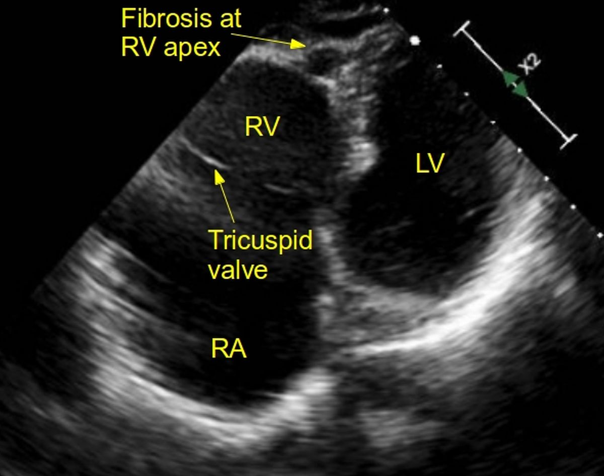
Echo image of right ventricular apex (RVA) pacemaker, pressure
Por um escritor misterioso
Last updated 22 março 2025


Figure. Exemplificative diagram for RVOT septal and RVAP pacing. Chest

Left Ventricular Pacing. Is It Always Better Than Right Ventricular Pacing?

Right ventricular lead apical and septal locations.
JCM, Free Full-Text

Impact of Diastolic Interventricular Septal Flattening on Clinical Outcome in Patients With Severe Tricuspid Regurgitation

Adverse effects of right ventricular pacing on cardiac function: prevalence, prevention and treatment with physiologic pacing - ScienceDirect
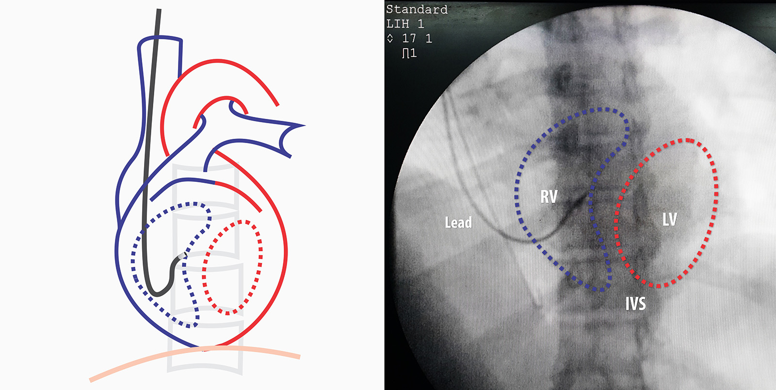
Septal Implantation of Right Ventricular Lead – How to Pace
Multiple echo views in EMF – All About Cardiovascular System and Disorders

Selective site right ventricular pacing

Echocardiography and Heart Failure: A Glimpse of the Right Heart - Pleister - 2015 - Echocardiography - Wiley Online Library

Advantage of Right Ventricular Outflow Tract Pacing on Cardiac Function and Coronary Circulation in Comparison with Right Ventricular Apex Pacing
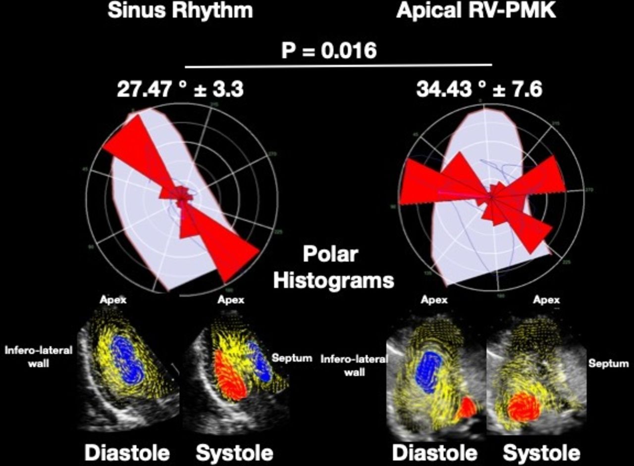
Intraventricular flow patterns during right ventricular apical pacing
The short term influence of right ventricular pacing burden on echocardiographic and spiroergometric parameters in patients with preserved left ventricular ejection fraction, BMC Cardiovascular Disorders

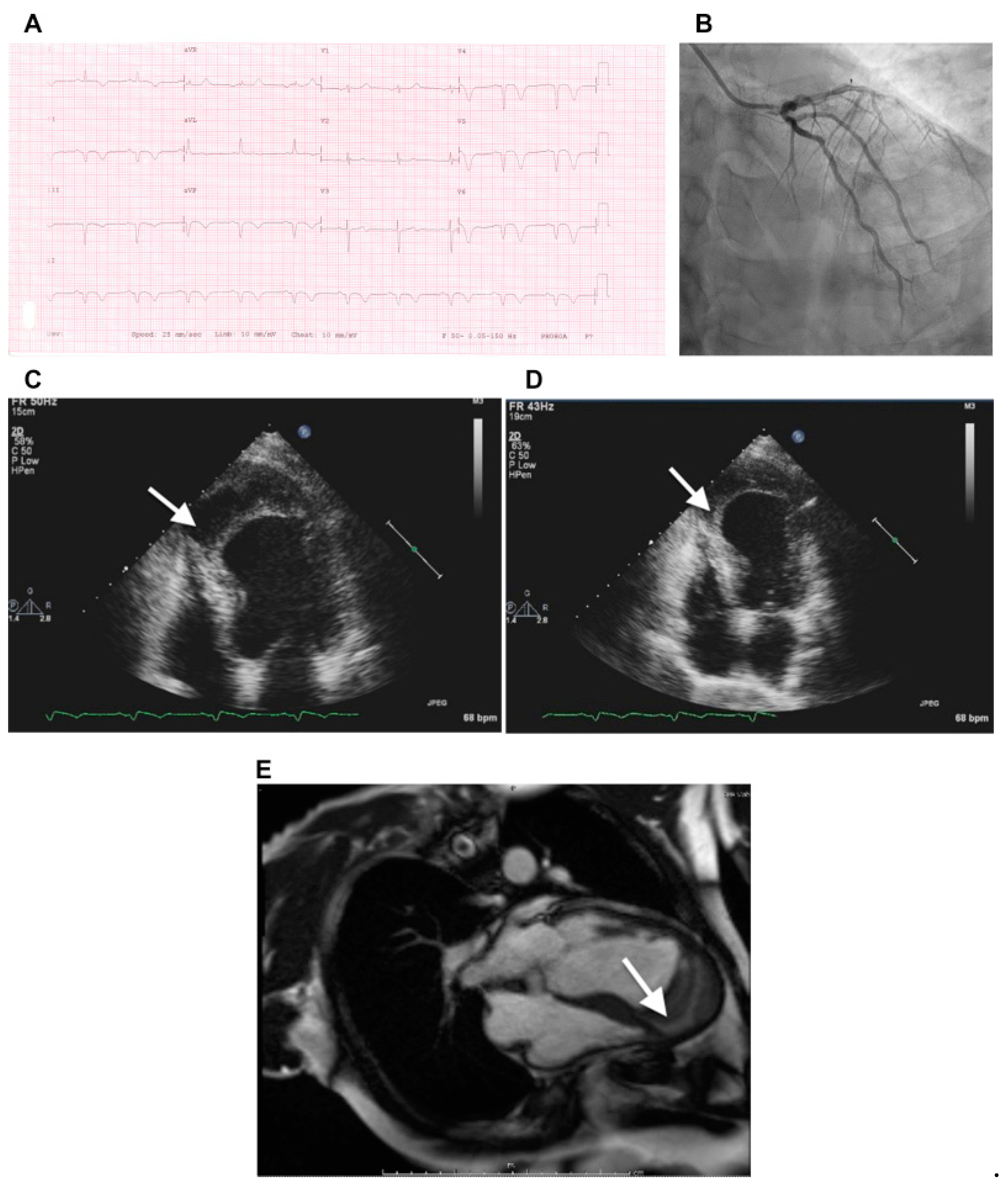
Delayed Right Ventricular Perforation by a Transvenous Active Fixation Implantable Cardioverter-Defibrillator Lead

Intraventricular flow patterns during right ventricular apical pacing
Recomendado para você
-
Radio RVA AM – Apps no Google Play22 março 2025
-
 Radio Venancio Aires - AM 910 - Venancio Aires, RS - Ouça Online22 março 2025
 -
 Typical Rapid Visco Analysis (RVA) profile of heat treated flour22 março 2025
 -
 Aposentadoria rural e segurança pública são os assuntos mais22 março 2025
 -
Mark Whitten will be at Anime Ink this October! Mark is the22 março 2025
-
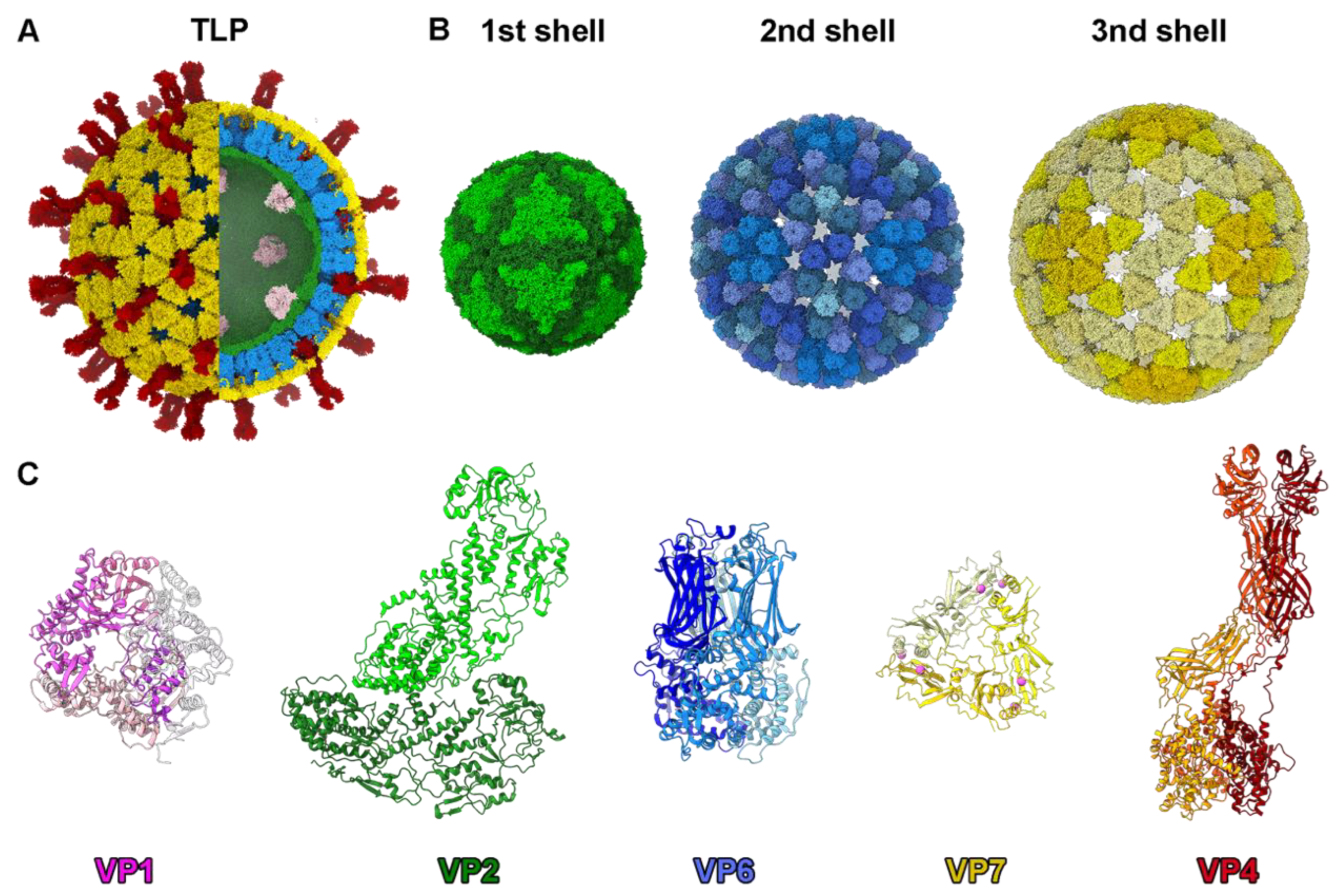 Viruses, Free Full-Text22 março 2025
 -
 Momentum Voya E+ 1 - Pedal Power RVA22 março 2025
 -
 15th annual RVA Latino Festival is here22 março 2025
 -
 The structure, properties and potential probiotic properties of starch-pectin blend: A review - ScienceDirect22 março 2025
 -
 Pesquisa Nupes/Unisc confirma RVA AM e Venus FM como líderes de22 março 2025


você pode gostar
-
 Call Me Tonight OVA (1986, ENG SUB)22 março 2025
 -
 Apartment carnon plage, Carnon-Plage, France22 março 2025
 -
 TABS】K-ON! S2 EP1 -「Yui's Solo」by @Tron54422 março 2025
 -
 eGames Expect More22 março 2025
 -
 Psychobob's Atlas Bot from Portal 2 3D Printed - 3D Printing Industry22 março 2025
 -
 Inter vs AC Milan: Complete H2H record in the Champions League22 março 2025
 -
:quality(70)/cloudfront-us-east-1.images.arcpublishing.com/metroworldnews/7RKCP53PKZF2HKLL23RTV23D3A.jpg) Esta é a série brasileira para maiores de 18 anos escondida na HBO22 março 2025
Esta é a série brasileira para maiores de 18 anos escondida na HBO22 março 2025 -
Livro Infantil 365 Atividades Barbie Para Pintar22 março 2025
-
 Light Novel - Volume 3, Arifureta Shokugyou de Sekai Saikyou Wiki22 março 2025
 -
 Tata Steel Masters: Four in the lead after Round 222 março 2025